We have unintentionally discovered a very simple way of telling whether
an MR solution is correct or not, provided that (as in this case) native
data have been measured to about 2.1A or better. This uses the current
beta-test of SHELXE that does autotracing (available on email request).
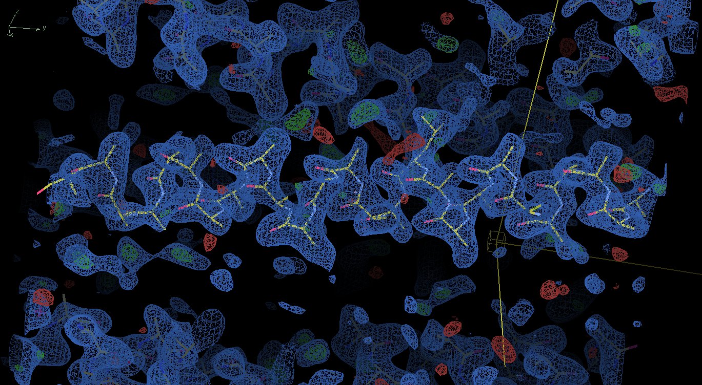
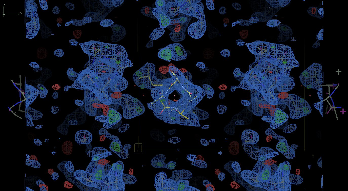
First rename the PDB file from MR to name.pda and generate a SHELX
format file name.hkl, e.g. using Tim Gruene's mtz2hkl, where 'name' may
be chosen freely but should be the same for both input files. Then run
SHELXE with a large number of autotracing cycles (here 50), e.g.

shelxe name.pda -a50 -s0.5 -y2

-s sets the solvent content and -y a resolution limit for generating
starting phases. If the .hkl file contains F rather than intensity the
-f switch is also required.

If the model is wrong the CC value for the trace will gradually
decrease as the model disintegrates. If the model is good the CC will
increase, and if it reaches 30% or better the structure is solved. In
cases with a poor but not entirely wrong starting fragment, the CC may
vary erratically for 10-30 cycles before it locks in to the correct
solution and the CC increases over three or four cycles to the value
for a solved structure (25 to 50%). The solution with the best CC is
written to name.pdb and its phases to name.phs for input to e.g. Coot.

George

On Fri, Nov 04, 2011 at 10:42:27AM +0100, Sergei Strelkov wrote:
> Dear Napoleão,
>
> Thank you for updating everyone on your
> efforts, and also acknowledging the advice.
>
> I wanted to respond to your question regarding maps.
> I know that many people who try to figure out
> whether or not their MR solution is the right one
> would ask the same question.
>
> So first of all if you wonder why you actually get very
> decently looking maps the answer is a classical one:
> because 'the phases are more important than amplitudes'.
> The appearance of your map is defined by
> your model phases, and hence a good match between the
> model and the map may not be taken as
> a sign of a correct solution. Once again: never ever!
>
> On the contrary, at least in your coil1.jpg
> image I clearly see that the density exactly follows
> the model which is almost entirely poly-Ala.
> Unless your protein is really poly-Ala this should be
> alarming. If you had a correct solution they you
> would hope to see the (difference)density for
> at least some missing side chains.
>
> And a second point. Unless your model contains
> the complete chain (which is rarely the case, especially
> for the coiled coils, as discussed already)
> a sign of the correct solution would be the appearance
> of extra density near the N- and/or C-terminus of
> the model. If it is not there, it is almost certainly not
> a solution.
>
> And you should not be worried about the R-factors being
> very high at this stage. If the solution is correct then
> you should see at least some extra features in the map.
>
> Kind regards,
> Sergei
>
>
>
>     Thank you all for the replies.
>
>     Sorry for taking so long to reply, I was actually trying some of your
>     interesting ideas (and I'm still trying).
>
>     I tried using the low resolution data sets for the molecular replacement
>     (thanks to Yuriy Patskovsky), I also improved and increased my coiled
>     coil database and employed it in many approaches using EPRM (interesting
>     program I was not aware of), which I found to produce lots of data,
>     hopefully addressing at some extent the helixes bent (thanks to Bernhard
>     Rupp). I also tried some more tweaking in Phaser, although not sure if
>     did it properly (thanks to Randy Read).
>
>     There is no twinning as far as I can tell (thanks to Ed Pozharski for
>     the tip). Using a data set with enough completeness (360 degrees @
>     Brookhaven) and processing in P1 did not help me because in this space
>     group there is most likely 2-3 helixes in the asymmetric unit, which
>     complicates the problem (and it takes a lot of time for Phaser to run).
>     Automated approaches also did not yield a better result (as far as I can
>     tell). I'm convinced that the space group is C2221, but I may be wrong.
>
>     Thanks to Sergei Strelkov for the numerous useful suggestions on how to
>     approach the problem.
>
>     One of the big issues for me is to discriminate between a lot of
>     similarly good density maps. For example:
>
>     http://www.fullonline.org/coils/coil1.jpg
>     http://www.fullonline.org/coils/coil2.jpg
>
>     I have hundreds of solutions like these and I think they are all wrong.
>
>     I couldn't manage to run Arcimboldo, could not find a tutorial on it
>     either. It was highly recommended here (and elsewhere), so I'm
>     definitely willing to give it a try (thanks Isabel Uson).
>
>     You guys opened my eyes about a series of issues that I should learn
>     about and approach, I'm most thankful for that.
>     Best regards,
>                       Napo
>
>
>
> --
> Prof. Sergei V. Strelkov
> Laboratory for Biocrystallography
> Department of Pharmaceutical Sciences, Katholieke Universiteit Leuven
> O&N2, Campus Gasthuisberg, Herestraat 49 bus 822, 3000 Leuven, Belgium
> Work phone: +32 16 330845  Fax: +32 16 323469 OR +32 16 323460
> Mobile:     +32 486 294132
> Lab pages: http://pharm.kuleuven.be/anafar
>

--
Prof. George M. Sheldrick FRS
Dept. Structural Chemistry,
University of Goettingen,
Tammannstr. 4,
D37077 Goettingen, Germany
Tel. +49-551-39-3021 or -3068
Fax. +49-551-39-22582